SPECT Imaging: A Window Into Brain Health
What Is SPECT Imaging and How Does It Help TBI Survivors?
SPECT (Single Photon Emission Computed Tomography) is a functional brain imaging technique that measures blood flow and metabolic activity across the brain, rather than just its physical structure. For traumatic brain injury (TBI) survivors, this matters because a SPECT scan can reveal areas of reduced or irregular blood flow even when a standard MRI or CT scan comes back "normal" — often giving survivors the first objective evidence of what they've been experiencing.
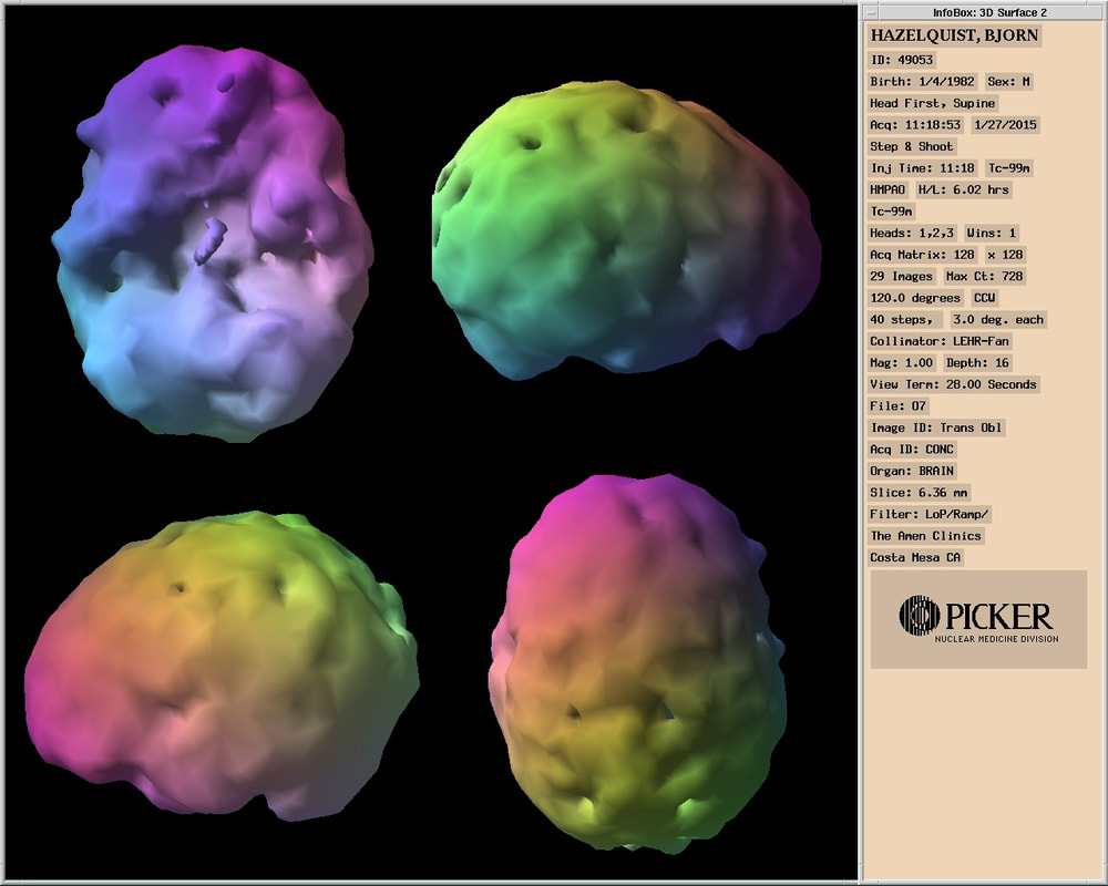
If you've been told your scans look fine but your symptoms say otherwise, that gap between imaging and experience is one of the most common — and most frustrating — parts of TBI recovery. SPECT imaging exists to help close it. The scan below is Bjorn's own, taken at the Amen Clinics in Costa Mesa in January 2015, after years of normal-looking CT and MRI results.
Why Traditional Scans Miss So Much
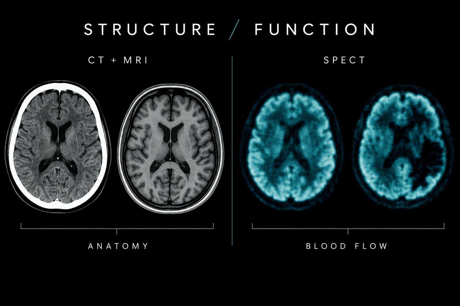
MRI and CT scans are built to answer a structural question: is there bleeding, a fracture, a visible lesion, swelling? They're essential for ruling out life-threatening injuries in the emergency room, and they remain the standard first step after any significant head trauma. But a large share of concussions and mild-to-moderate TBIs never produce a structural abnormality that these scans can catch. The brain can be functionally injured — its blood flow and metabolism disrupted — without a single visible mark on an MRI or CT image.
This is part of why TBI misdiagnosis is so common in the first place. Structural scans simply weren't designed to detect functional injury, which leaves many survivors with normal imaging and persistent, real symptoms that no one can explain.
How SPECT Imaging Works
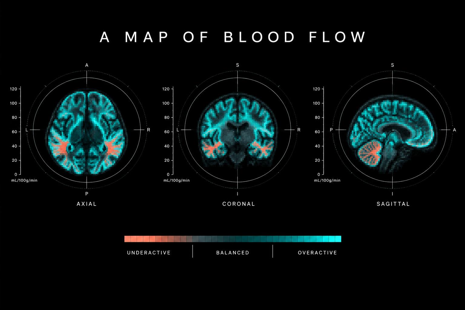
SPECT imaging uses a small amount of a radioactive tracer, injected into the bloodstream, that travels to the brain and emits gamma rays as it circulates. A specialized camera detects this activity and builds a three-dimensional map showing which regions of the brain are receiving strong blood flow, which are underactive, and which show patterns associated with injury. Areas of the brain working overtime — often linked to anxiety, hypervigilance, or compensatory strain — can show up as overactive. Areas struggling to recover from injury frequently show up as underactive, or hypoperfused.
Unlike MRI, which shows anatomy, or an EEG, which measures electrical activity, SPECT is a perfusion study — it's specifically mapping blood flow. That distinction is what allows it to pick up functional disruption that structural scans pass right over.
What the Research Shows
SPECT imaging for TBI isn't a new idea — it dates back to techniques developed in the 1960s and has since been studied extensively in patients with head injury, cognitive impairment, and psychiatric conditions that commonly co-occur with brain trauma. A systematic review of decades of SPECT-TBI research found that in the large majority of comparison studies, SPECT identified lesions or areas of dysfunction that structural CT and MRI scans either missed entirely or significantly understated in size. In studies that went a step further and correlated SPECT findings with neuropsychological testing, a strong majority found a statistically significant relationship between the blood flow abnormalities SPECT visualized and the cognitive deficits patients were actually experiencing.
Researchers have also identified recurring patterns of reduced blood flow — or hypometabolism — in specific brain regions among people with mild TBI and lingering, persistent symptoms, including the posterior cingulate gyrus, the parieto-occipital and frontal lobes, the temporal lobe, and the cerebellum. That consistency across studies is part of what has made SPECT a serious research tool in this space, not just an experimental curiosity.
None of this means SPECT is perfect. The scientific literature acknowledges genuine disagreement in places, and SPECT produces lower-resolution images than PET or fMRI. But its lower cost, wider availability, and ability to flag functional injury that structural imaging misses have kept it in active clinical and research use for TBI, cognitive disorders, and cerebrovascular disease alike.
From Imaging to Individualized Treatment
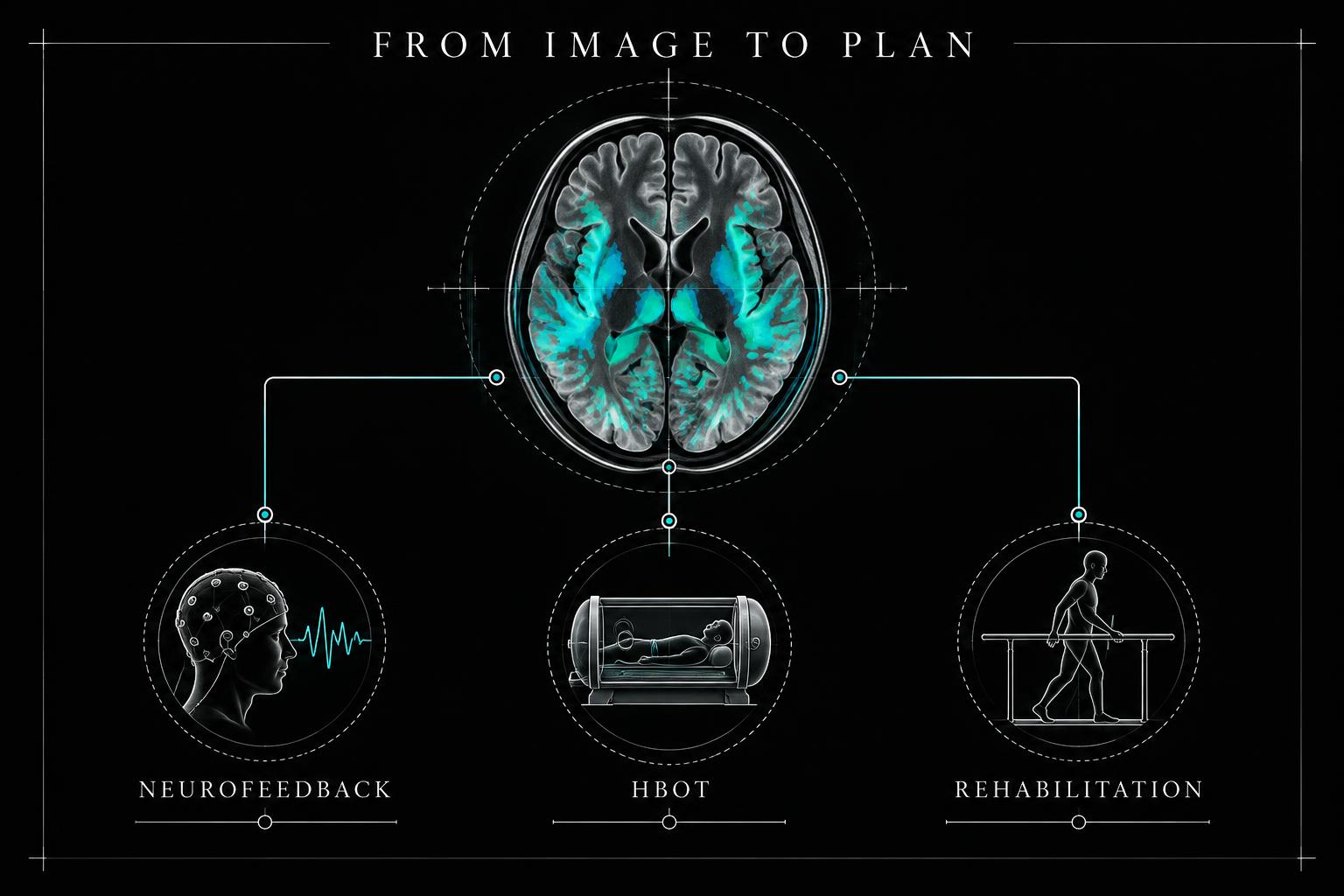
A picture of dysfunction is only useful if it changes what happens next — and that's where SPECT earns its place in a TBI recovery plan. By showing clinicians which specific regions of the brain are underperforming or overactive, a SPECT scan can help guide decisions that would otherwise be made through trial and error: which type of therapy might target which symptom cluster, whether a treatment like neurofeedback or hyperbaric oxygen therapy (HBOT) is worth pursuing, and how to track whether an intervention is actually changing brain function over time, not just symptoms on a questionnaire.
For survivors who have spent months or years being told their scans are normal, that specificity can be transformative — turning a vague, dismissed injury into a mapped, addressable one, and turning a generic treatment plan into one built around their actual brain.
Who Might Benefit From a SPECT Scan
SPECT imaging tends to be most useful for TBI survivors who:
• Have persistent cognitive, mood, sleep, or physical symptoms months after injury
• Received "normal" results on a CT or MRI despite ongoing symptoms
• Are struggling to get a diagnosis taken seriously by providers or insurers
• Want objective data to guide decisions about neurofeedback, HBOT, or other emerging therapies
• Have overlapping symptoms of TBI, anxiety, or PTSD and need help distinguishing what's driving what
It isn't a first-line emergency tool, and it isn't necessary for every survivor. But for the large population of people stuck in the gap between "my scans are clear" and "something is clearly wrong," it can be one of the most clarifying steps in a recovery journey.
What to Expect During a SPECT Scan
The process is more accessible than many people expect. A technician administers the tracer through an IV, and after a short uptake period, the patient lies still on a table while the SPECT camera rotates around the head, capturing images from multiple angles. The scan itself typically takes 15 to 30 minutes and is not painful. Radiation exposure is low and comparable to other standard nuclear medicine imaging. Results are then reviewed by a physician trained in interpreting brain perfusion patterns, who translates the imaging into practical findings a survivor and their care team can actually act on.
Accessing SPECT Imaging Through the Foundation
Because SPECT imaging isn't always covered by insurance and can be difficult to access without the right referrals, Strength in Pain Foundation offers guidance and financial assistance to help survivors pursue this technology when it's medically appropriate. If you've been struggling with symptoms that never showed up on a traditional scan, SPECT imaging may offer the answers — and the direction — you've been looking for. Contact us to learn more about our financial assistance programs and how to get started.
Frequently Asked Questions
Does SPECT imaging show things MRI and CT scans miss?
Yes. MRI and CT scans look at brain structure — bleeding, fractures, visible lesions. SPECT measures brain function through blood flow, which means it can pick up on injury-related dysfunction even when structural scans appear completely normal.
Is SPECT imaging safe?
Generally, yes. It uses a low dose of a radioactive tracer, similar to other standard nuclear medicine procedures, and the scan itself is non-invasive and painless.
How long does a SPECT scan take?
The imaging portion typically takes 15 to 30 minutes, though there's usually a waiting period after tracer injection before the scan begins.
Will insurance cover a SPECT scan for a brain injury?
Coverage varies widely by insurer and by the medical necessity documented for the scan. Many TBI survivors face out-of-pocket costs, which is part of why the Foundation offers financial assistance for this and other emerging therapies.
Can SPECT imaging diagnose a TBI on its own?
SPECT is best used alongside a full clinical evaluation — history, symptoms, and other testing — rather than as a standalone diagnostic tool. Its greatest value is in confirming and localizing functional injury that other tests can't see, and in helping guide a personalized treatment plan.
This article is educational and is not medical advice. Always consult a qualified healthcare provider about your specific symptoms and treatment options. If you are experiencing a medical emergency, call 911.